Lateral/Lower Eyelid Reconstruction Reviews View More
SISUN PREMIUM EYE SURGERY

Lateral Canthoplasty Reconstruction

Correct unnatural outer-corner contours, scarring, and exposed mucosa after lateral canthoplasty.

  • Surgery Duration
    30 min
  • Anesthesia
    IV Sedation / Local
  • Stitch Removal
    10 days
  • Hospitalization
    Same-day Discharge
  • Recovery Period
    7~10 days

*Individual results may vary. Side effects such as bleeding, infection, and inflammation may occur after cosmetic surgery. Please consult your doctor.

Lateral Canthoplasty Reconstruction — What is it?

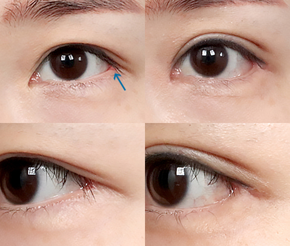
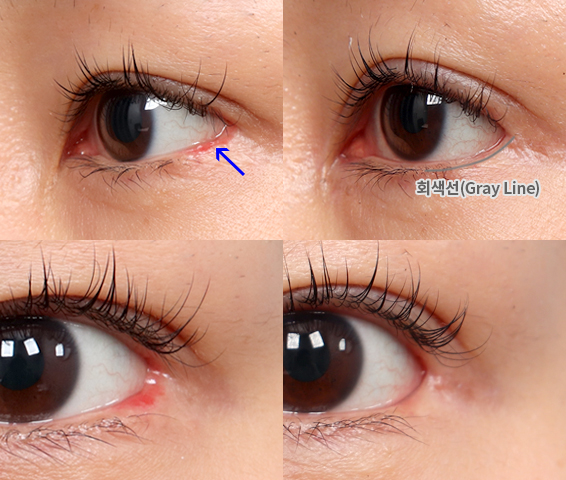
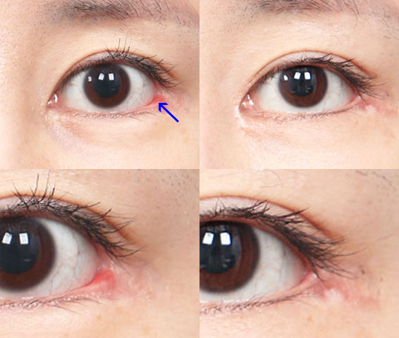
  • TYPE 01

    Square/C shape

  • TYPE 02

    Red scar (conjunctival exposure)

  • TYPE 03

    Unnatural line

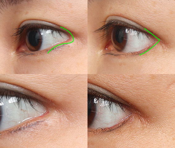
If the outer corner has become squared off or C-shaped, red scarring is visible because of conjunctival exposure, or the contour is uneven, the ligament and conjunctival layers are repositioned to improve the scar and exposed mucosa.

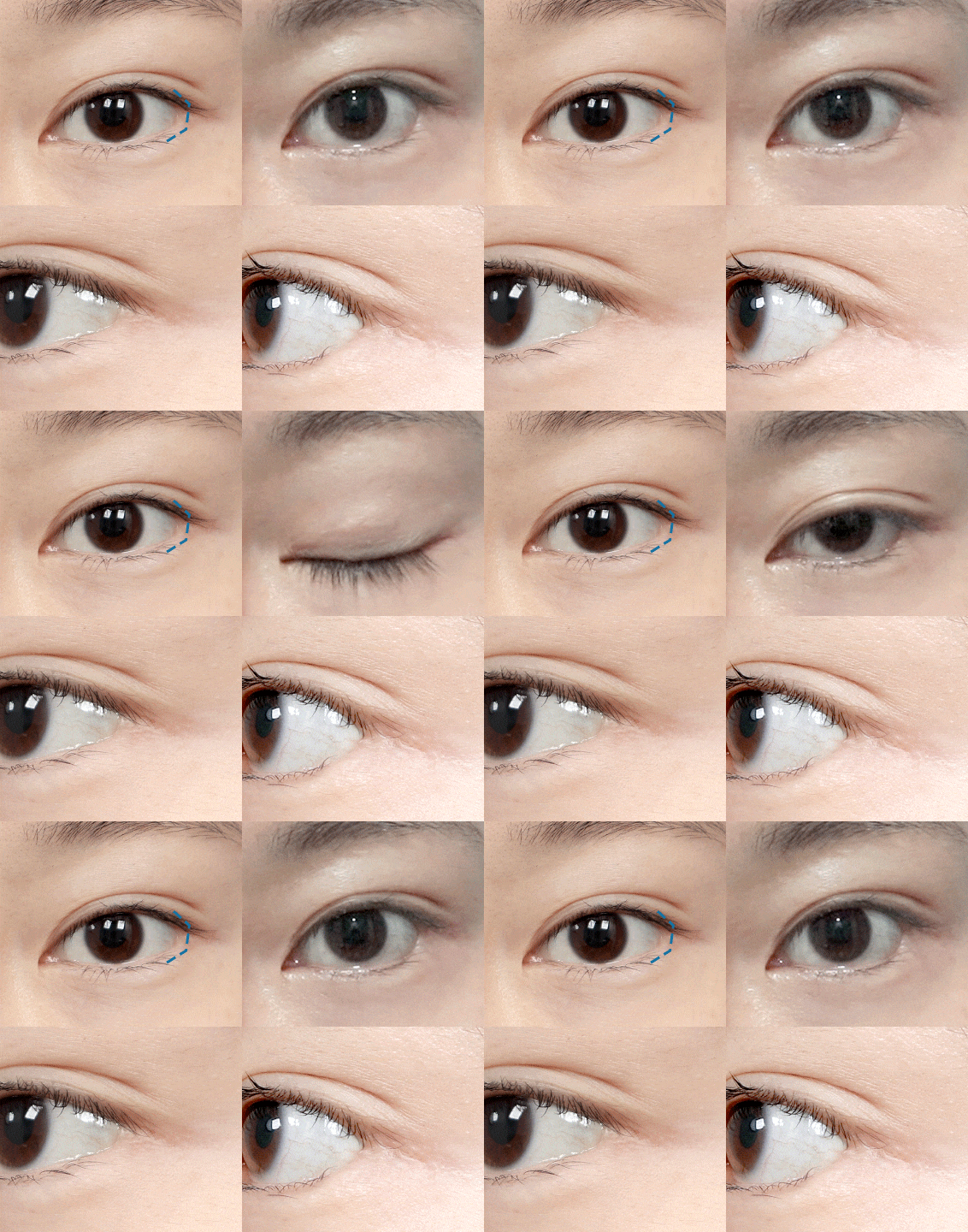
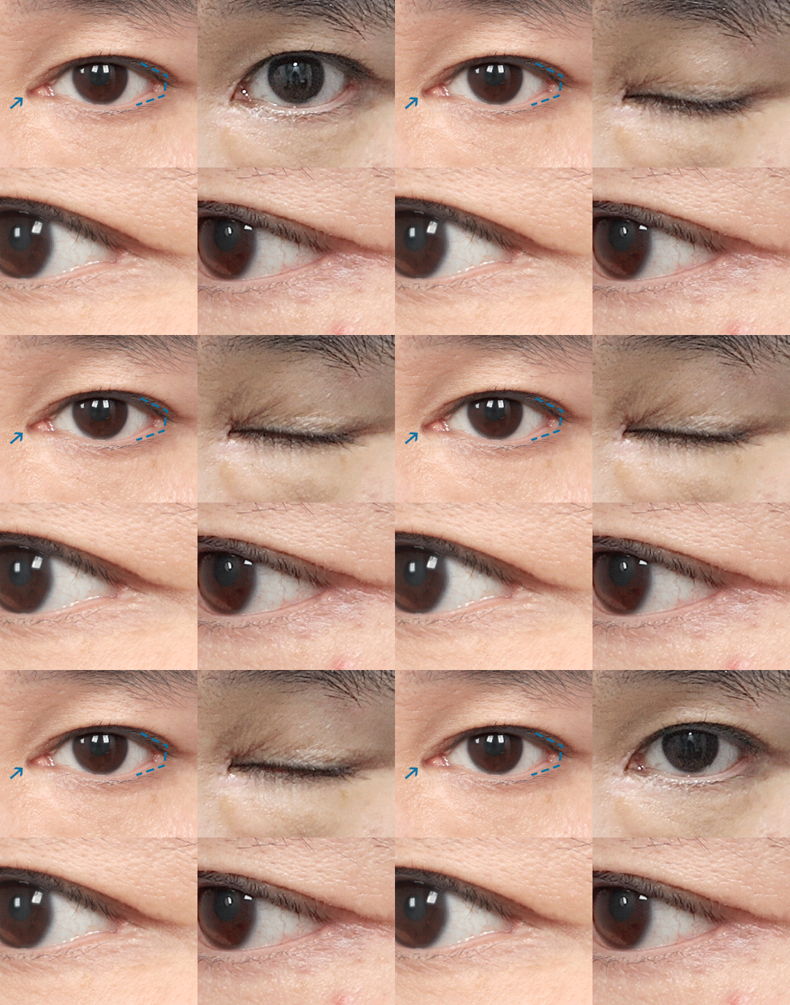
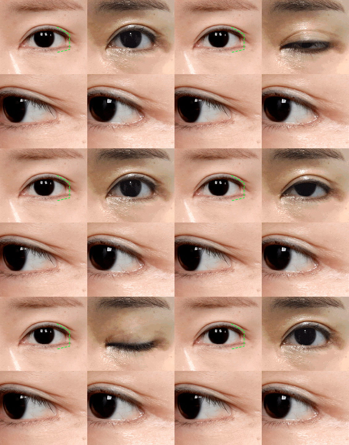
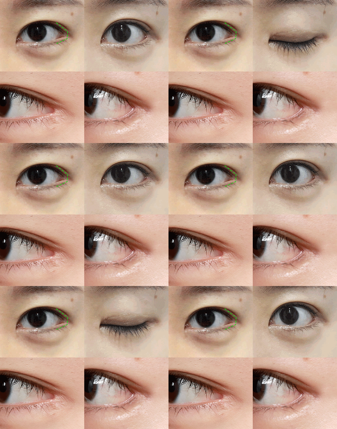
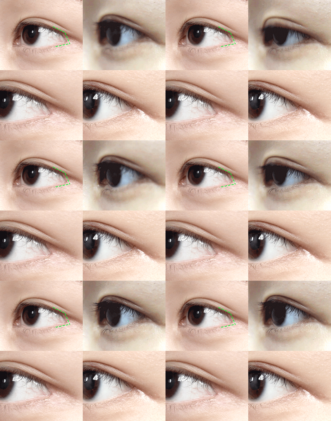
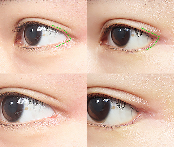
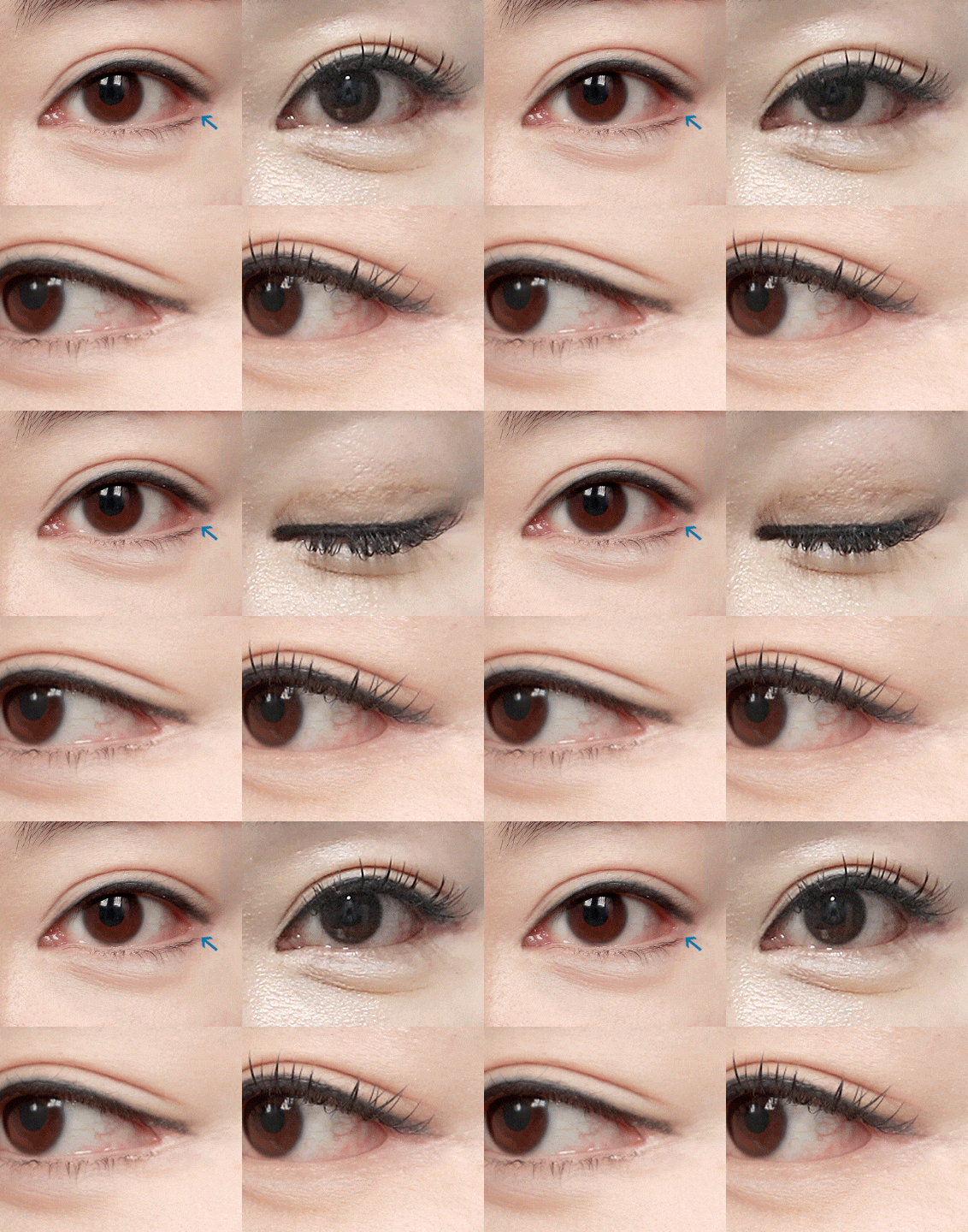
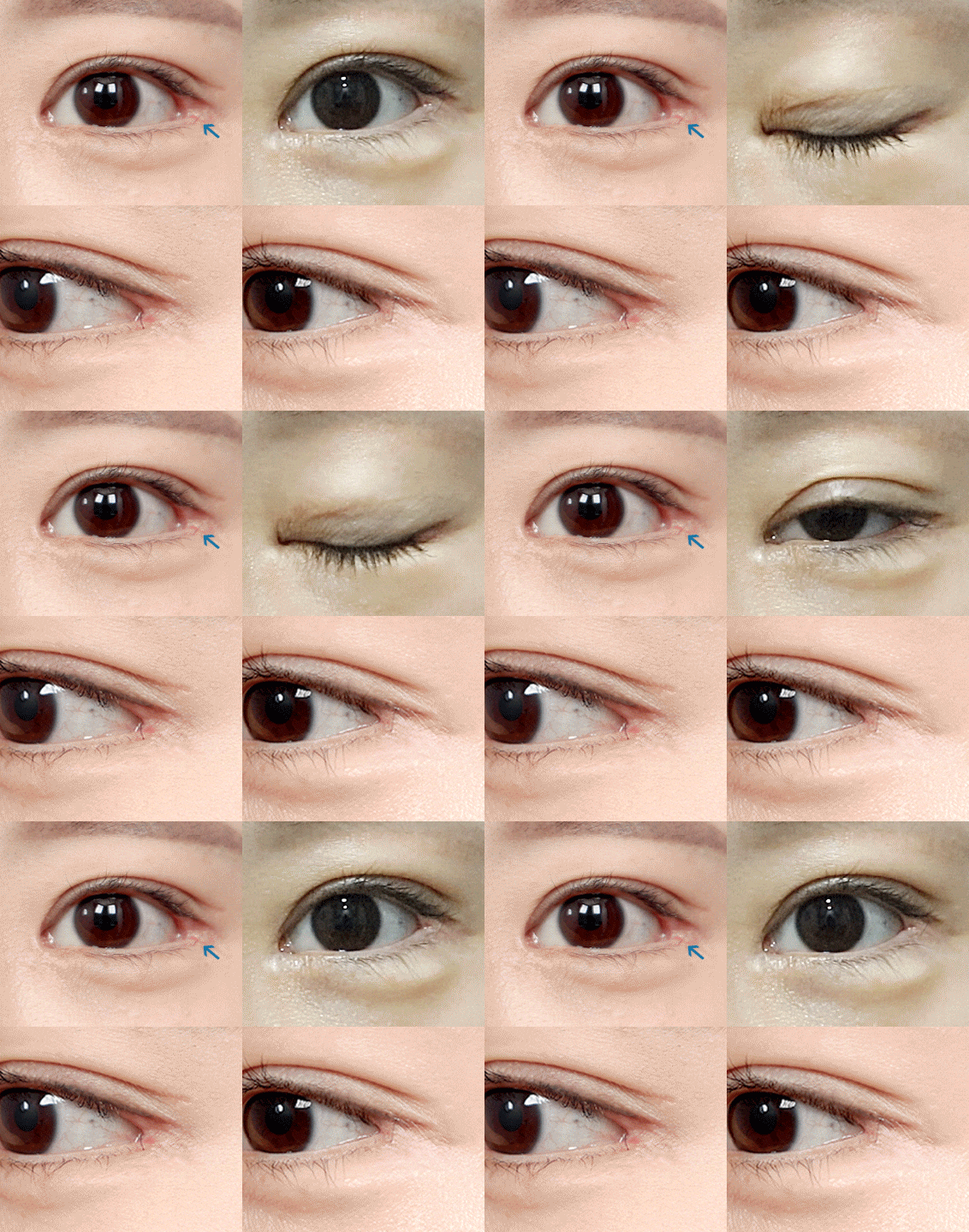
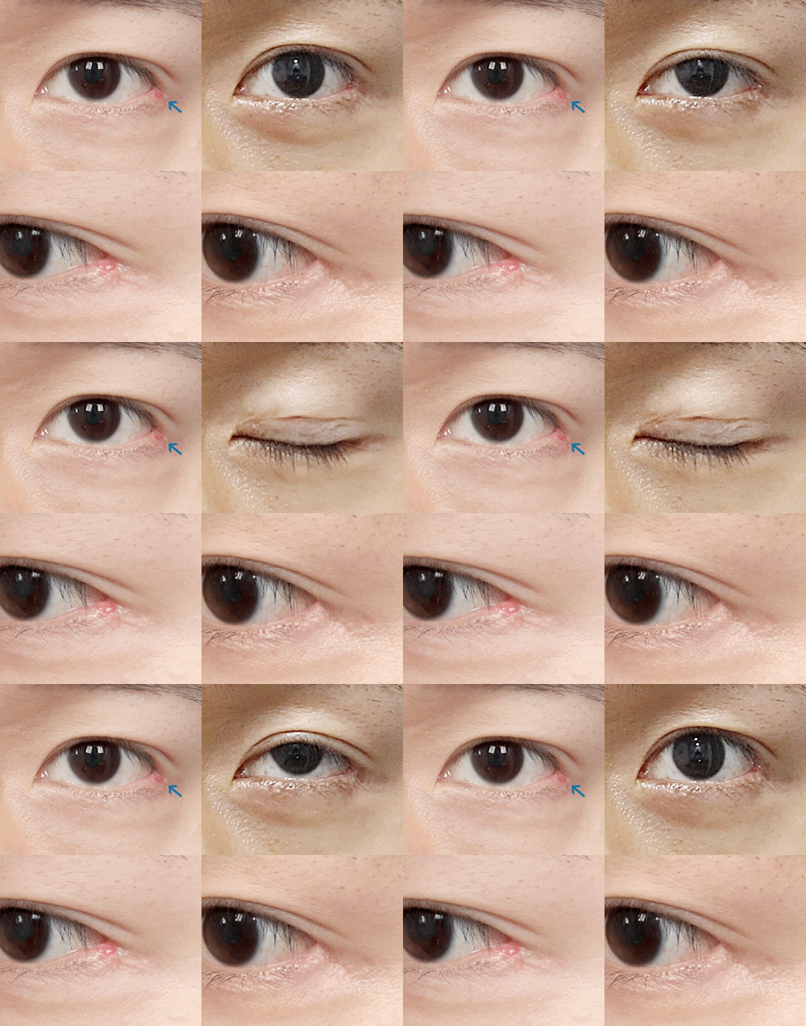
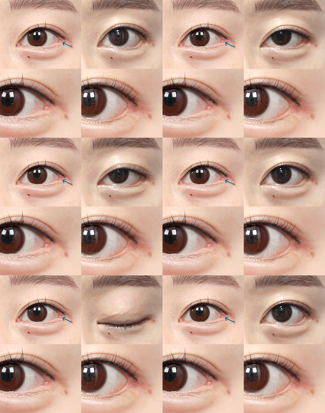
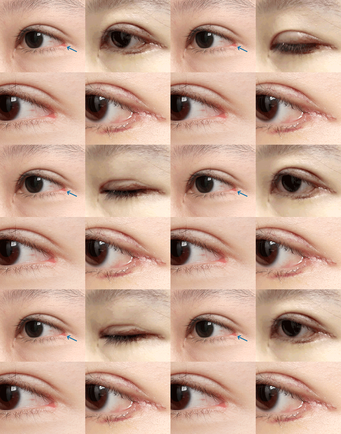
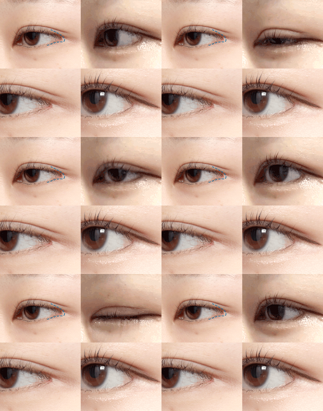
  • Lateral Canthoplasty Reconstruction+Lower Eyelid Surgery (7 weeks)

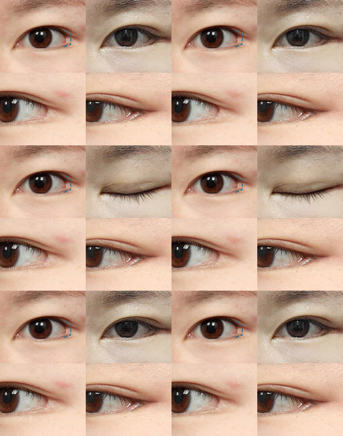
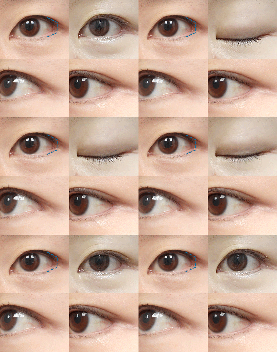
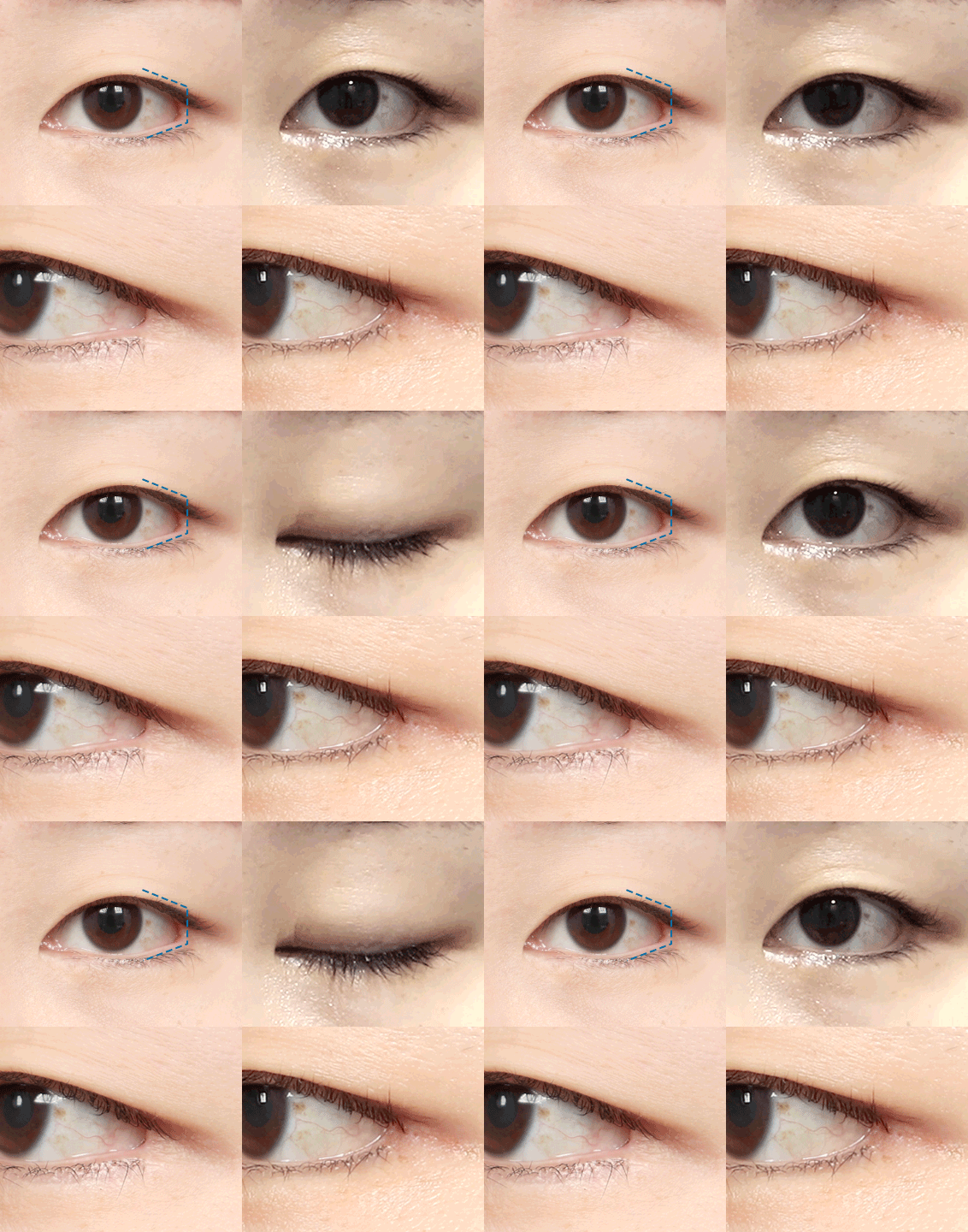
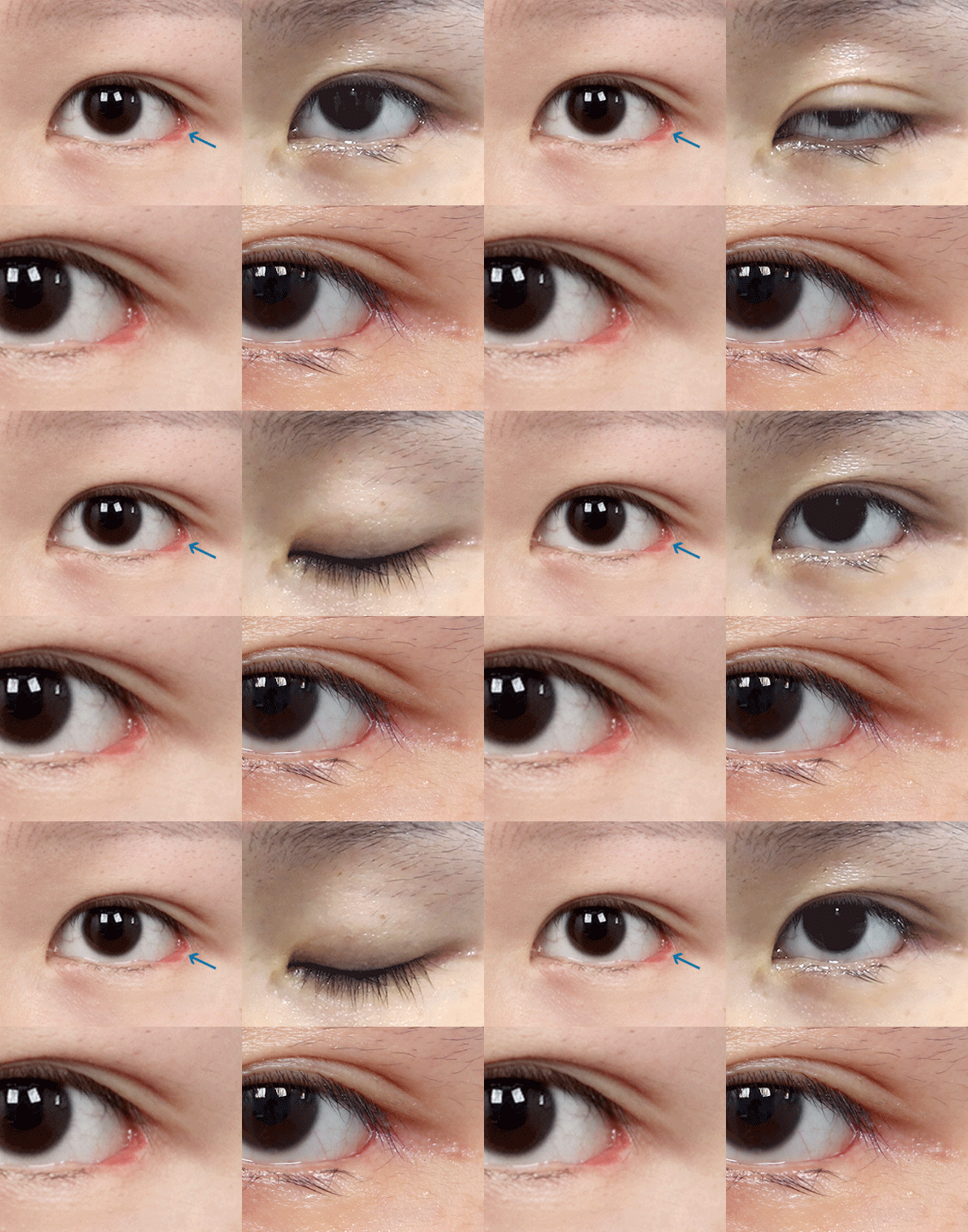
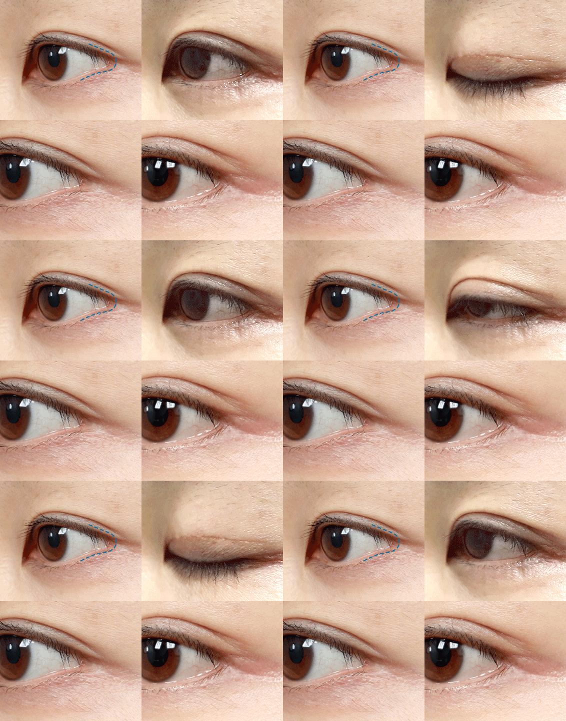
  • Lateral Canthoplasty Reconstruction+Lower Eyelid Surgery (revision) (5 weeks)

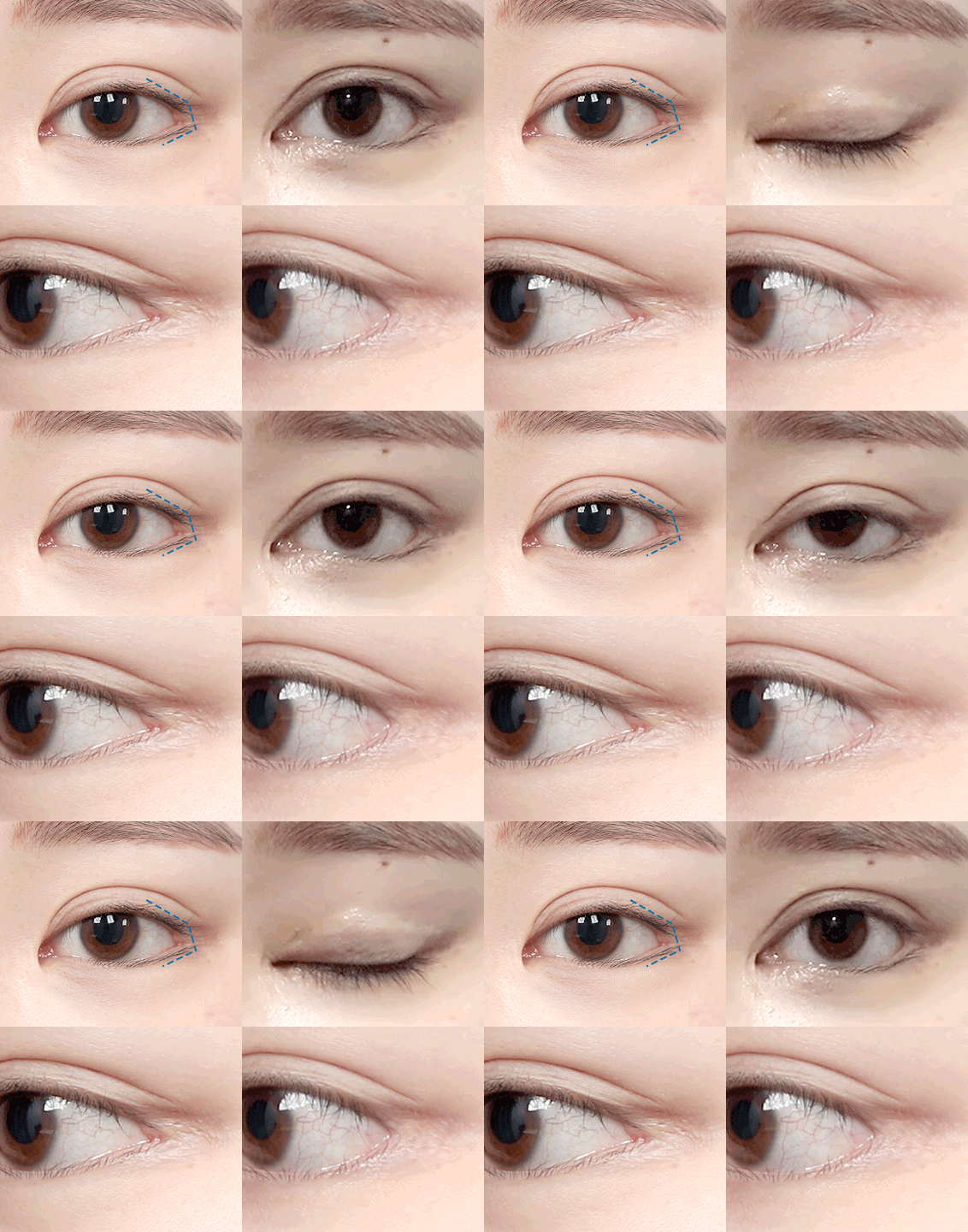
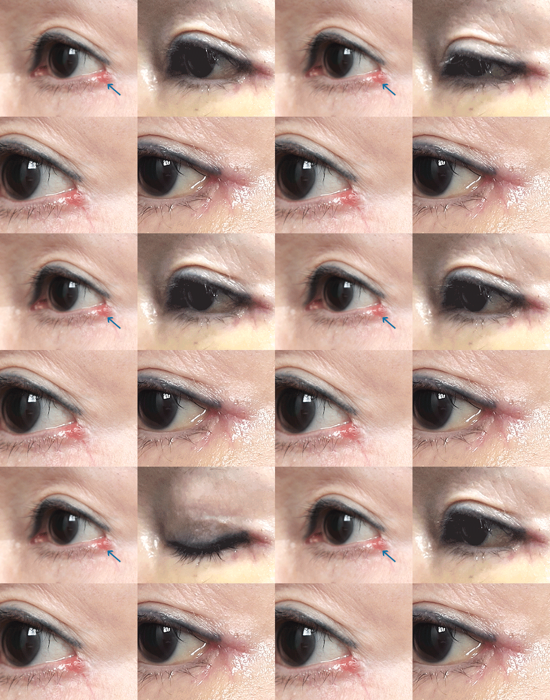
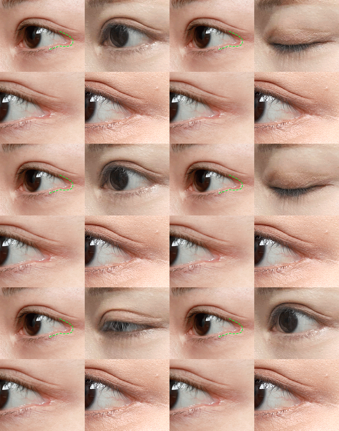
  • Lateral Canthoplasty Reconstruction+Lower Eyelid Surgery (revision) (2 months)

Lateral Canthoplasty Reconstruction Surgical Candidates

  • Outer eye corners
    have an unnatural
    square or
    "C" shape
  • Noticeable
    scarring
  • Red conjunctiva
    is exposed
  • Dryness, pain,
    redness, inability
    to close eyes after
    lateral canthoplasty
  • Mucosa line
    has separated
    from the eyeball

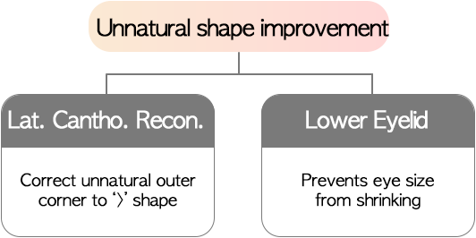
Lateral Canthoplasty Reconstruction, Why lower canthoplasty is more effective when combined with surgery

Will my eyes become smaller after Lateral Canthoplasty Reconstruction?

Yes, they can appear smaller.
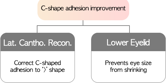
However, if lower eyelid surgery is performed at the same time,
it can help prevent that effect.

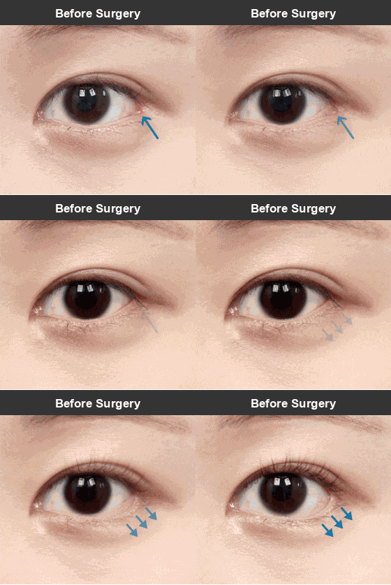
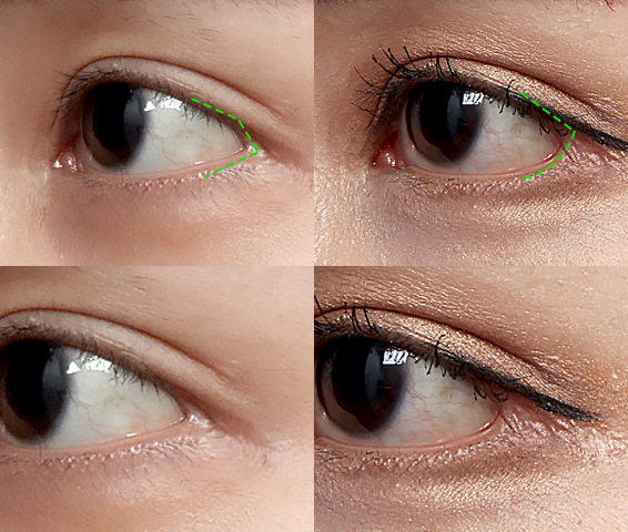
  • Conjunctival exposure, square adhesion

  • Lateral Canthoplasty Reconstruction

  • Lateral Canthoplasty Reconstruction+Lower Eyelid Surgery

The primary goal of Lateral Canthoplasty Reconstruction is to restore the eyes as close to their pre-surgery state as possible. Many patients who had lateral canthoplasty
hoping for bigger eyes but ended up with minimal results and scars, or unnatural outer corner lines,
say they want to be restored to their pre-surgery state even if their eyes become smaller again.

However, once the reconstruction is done, some patients feel frustrated by the smaller eye size and consider
lateral canthoplasty revision again. To prevent this, it is recommended to combine Lateral Canthoplasty Reconstruction with lower eyelid surgery.
This way, scars can be improved while preventing dissatisfaction from reduced eye size.

Lateral Canthoplasty Reconstruction Surgical Technique

Before Surgery

Outer eye corner has a square shape
with exposed mucosa

STEP01

Excise scar tissue and
exposed mucosa at the outer eye corner

STEP02

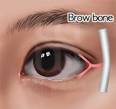
Pull the lateral canthal tendon
to the brow bone
and fix with sutures

STEP03

Adding lower eyelid surgery
can prevent the eyes
from becoming smaller

After Surgery

Restore a pointed outer-corner contour and address scarring and exposed mucosa

Lateral Canthoplasty Reconstruction Surgical Technique

Lateral Canthoplasty Reconstruction
Surgical Cases

01

Square adhesion

Case 1
Lateral Canthoplasty Reconstruction+Lower Eyelid Surgery

After lateral canthoplasty, both sides developed square-shaped adhesions
and the lower eyelid mucosa line separated from the eyeball, exposing red flesh.

1 week later

Although bruising and swelling are present, you can see the outer eye corner shape has changed.

6 weeks later

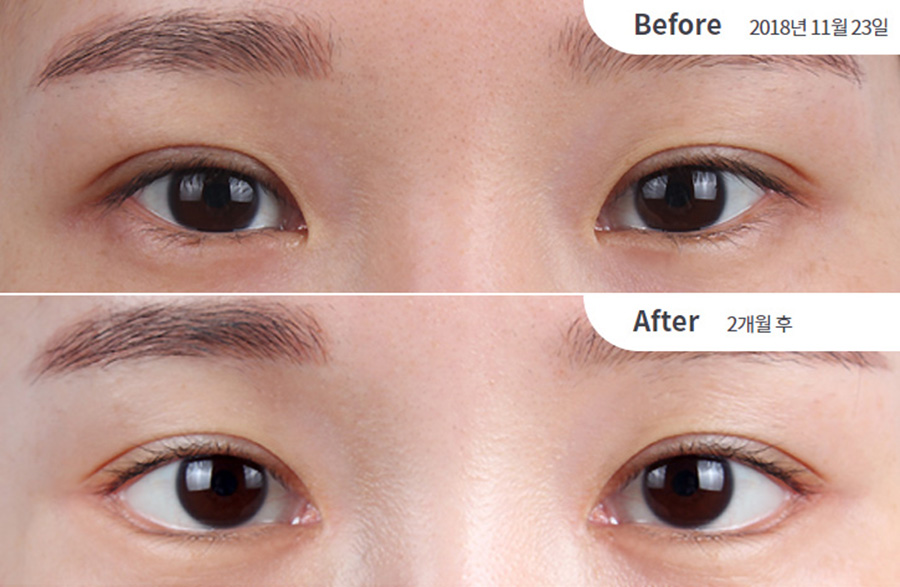
The square shape at the outer eye corner has been corrected without the eye size decreasing.

  • Before Surgery

  • 1 week later

  • 6 weeks later

Case 2
Lateral Canthoplasty Reconstruction+Lower Eyelid Surgery (revision)

After lateral canthoplasty, the left side developed a square scar and the right side had a torn-looking scar below the outer eye corner.
In such cases, a lateral canthoplasty revision would not improve the scars, so Lateral Canthoplasty Reconstruction must be performed.

1 week later

There is almost no swelling, but conjunctival edema has developed.

6 weeks later

The conjunctival edema has resolved and everything has healed well overall.

  • Before Surgery

  • 1 week later

  • 6 weeks later

  • Lateral Canthoplasty Reconstruction+Lower Eyelid Surgery (revision) (4 months)

  • Lateral Canthoplasty Reconstruction+Lower Eyelid Surgery (revision) (4 months)

  • Lateral Canthoplasty Reconstruction+Lower Eyelid Reconstruction (6 months)

  • Lateral Canthoplasty Reconstruction+Lower Eyelid Reconstruction (6 months)

  • Lateral Canthoplasty Reconstruction (1 year)

  • Lateral Canthoplasty Reconstruction (1 year)

  • Lateral Canthoplasty Reconstruction+Lower Eyelid Surgery (revision) (7 months)

  • Lateral Canthoplasty Reconstruction+Lower Eyelid Surgery (revision) (7 months)

  • Incisional Ptosis Correction (revision)+Epicanthoplasty Reconstruction+Upper Epicanthoplasty+Lateral Canthoplasty Reconstruction+Lower Eyelid Surgery (revision) (2 months)

  • Incisional Ptosis Correction (revision)+Epicanthoplasty Reconstruction+Upper Epicanthoplasty+Lateral Canthoplasty Reconstruction+Lower Eyelid Surgery (revision) (2 months)

  • Lateral Canthoplasty Reconstruction+Lower Eyelid Revision (Loosening)+Lower Eyelid Surgery (revision) (2 months)

  • Lateral Canthoplasty Reconstruction+Lower Eyelid Revision (Loosening)+Lower Eyelid Surgery (revision) (2 months)

  • 02

    Conjunctival exposure (red scar)

    Case 1
    Lateral Canthoplasty Reconstruction+Lower Eyelid Surgery

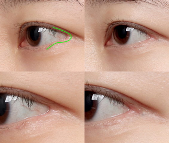
    After lateral canthoplasty, the conjunctiva was exposed on both sides
    and adhered in an unnatural C-shape.

    1 week later

    Although bruising and swelling remain, the outer eye corner shape has been corrected
    and the red scar has been removed.

    1 month later

    As it settled, the red scar is no longer visible
    and the pain felt when opening and closing the eyes has also disappeared.

    • Before Surgery

    • 1 week later

    • 1 month later

    Case 2
    Lateral Canthoplasty Reconstruction+Lower Eyelid Surgery (revision)

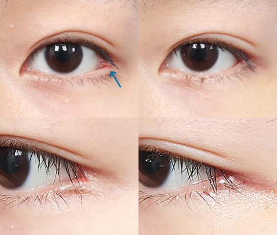
    After lateral canthoplasty, the conjunctiva was exposed on both sides, revealing red scars.
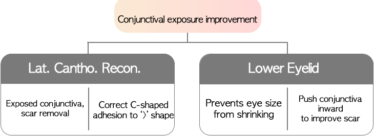
    Normally, both areas 1 and 2 where the conjunctiva is exposed should be removed,
    but this could make the eyes too small. So area 1 is restored,
    while area 2 has the exposed conjunctiva pushed inward with lower eyelid surgery to improve the scar.

    1 week later

    Upper epicanthoplasty, Lateral Canthoplasty Reconstruction, and lower eyelid revision were performed together,
    so significant swelling and scarring still remain.

    4 months later

    The red scar has been improved to be nearly invisible.

    • Before Surgery

    • 1 week later

    • 4 months later

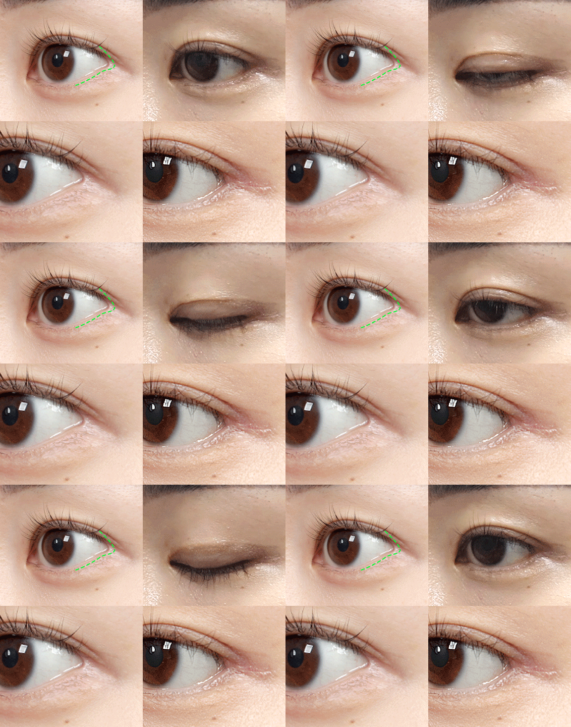
  • Lateral Canthoplasty Reconstruction+Lower Eyelid Surgery (revision) (1 year)

  • Lateral Canthoplasty Reconstruction+Lower Eyelid Surgery (revision) (1 year)

  • Lateral Canthoplasty Reconstruction+Lower Eyelid Surgery (revision) (5 weeks)

  • Lateral Canthoplasty Reconstruction+Lower Eyelid Surgery (revision) (5 weeks)

  • Lateral Canthoplasty Reconstruction (6 weeks)

  • Lateral Canthoplasty Reconstruction (6 weeks)

  • Lateral Canthoplasty Reconstruction (6 months)

  • Lateral Canthoplasty Reconstruction (6 months)

  • Lateral Canthoplasty Reconstruction (revision) (1 year)

  • Lateral Canthoplasty Reconstruction (revision) (1 year)

  • Lateral Canthoplasty Reconstruction+Lower Eyelid Surgery (revision) (3 years 4 months)

  • Lateral Canthoplasty Reconstruction+Lower Eyelid Surgery (revision) (3 years 4 months)

  • Lateral Canthoplasty Reconstruction+Lower Eyelid Surgery (revision) (1 year 1 month)

  • Lateral Canthoplasty Reconstruction+Lower Eyelid Surgery (revision) (1 year 1 month)

  • Lateral Canthoplasty Reconstruction (4 months)

  • Lateral Canthoplasty Reconstruction (4 months)

  • Incisional Ptosis Correction (revision)(Crease Lowering)+Epicanthoplasty Reconstruction+Lateral Canthoplasty Reconstruction (1 year 7 months)

  • Incisional Ptosis Correction (revision)(Crease Lowering)+Epicanthoplasty Reconstruction+Lateral Canthoplasty Reconstruction (1 year 7 months)

  • 03

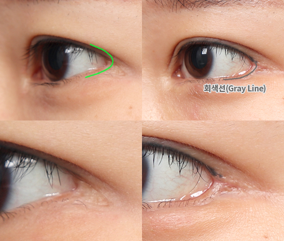
    Conjunctival separation (gray line damage)

    Case 1
    Lateral Canthoplasty Reconstruction+Lower Eyelid Surgery

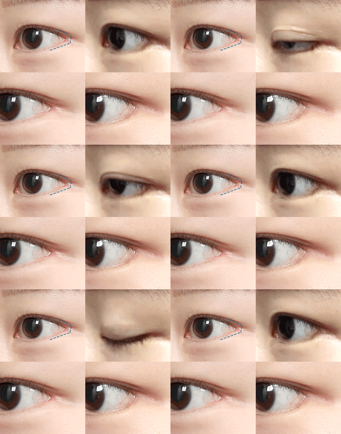
    After lateral canthoplasty, the left side had an unnatural square shape with red scars,
    and the right side had severe conjunctival exposure with red scars.

    1 week later

    Fine incision scars and redness remain, but the outer eye corner shape has been corrected.

    3 months later

    The incision scar is not noticeable, and the previously deformed
    outer eye corner shape has been improved to a pointed ">" shape.

    • Before Surgery

    • 1 month later

    • 3 months later

  • Lateral Canthoplasty Reconstruction+Lower Eyelid Surgery (revision) (2 months)

  • Lateral Canthoplasty Reconstruction+Lower Eyelid Surgery (revision) (2 months)

  • Lateral Canthoplasty Reconstruction+Lower Eyelid Surgery (revision) (1 year 1 month)

  • Lateral Canthoplasty Reconstruction+Lower Eyelid Surgery (revision) (1 year 1 month)

  • Lateral Canthoplasty Reconstruction (1 month)

  • Lateral Canthoplasty Reconstruction (1 month)

  • Lateral Canthoplasty Reconstruction+Lower Eyelid Surgery (revision) (7 months)

  • Lateral Canthoplasty Reconstruction+Lower Eyelid Surgery (revision) (7 months)

  • Lateral Canthoplasty Reconstruction+Lower Eyelid Surgery (revision) (3 months)

  • Lateral Canthoplasty Reconstruction+Lower Eyelid Surgery (revision) (3 months)

  • Lateral Canthoplasty Reconstruction+Lower Eyelid Surgery (3 years)

  • Lateral Canthoplasty Reconstruction+Lower Eyelid Surgery (3 years)

  • Lateral Canthoplasty Reconstruction

    Select photos with a Click badge to read the full case on our blog.
    01Square / C shape
    Case 7
    뒤트임복원+밑트임(재)(6주)
    Before
    After
    Assessment

    The outer eye corner was unnaturally bent and over-opened, causing ocular irritation.

    Procedure Notes

    The over-opened outer corner was reconstructed into a defined pointed shape, while lower canthoplasty refined the lower eye line into a rounded contour. This also helped prevent a reduction in visible eye size.

    Case 8
    뒤트임복원+밑트임(재)(2개월)
    Before
    After
    Assessment

    Previous poorly performed lateral and lower canthoplasty had left the outer corner adhered in an unnatural squared shape.

    Procedure Notes

    With lateral canthoplasty reconstruction and lower-canthoplasty revision, the previously squared, unnatural outer corner was corrected into a natural rounded shape.

    Case 9
    뒤트임복원+밑트임(재)(2개월)
    Before
    After
    Assessment

    A lateral-canthoplasty complication had left the outer corner separated, with the outer eye area appearing squared.

    Procedure Notes

    The separated squared outer corner was reconstructed into a defined pointed contour, and lower canthoplasty was performed at the same time to help prevent reduction of visible eye size, resulting in a more natural eye shape.

    Case 10
    뒤트임복원(1주)
    Before
    After
    Assessment

    A poorly performed lateral canthoplasty two years earlier had left the outer corner adherent in a squared shape, creating an unnatural appearance.

    Procedure Notes

    Lateral canthoplasty reconstruction corrected the squared shape into a defined, pointed outer corner.

    Case 11
    뒤트임복원+밑트임(재)(1주)
    Before
    After
    Assessment

    After lateral and lower canthoplasty one year earlier, the outer corner had adhered in a squared shape, with lower-lid line irregularity and scarring.

    Procedure Notes

    Lateral canthoplasty reconstruction removed the scar and refined the outer corner into a pointed contour; lower-canthoplasty revision helped prevent a reduction in visible eye size.

    Case 12
    뒤트임복원+밑트임(재)(1개월)
    Before
    After
    Assessment

    Following lateral and lower canthoplasty four years earlier, the outer corner had a squared shape.

    Procedure Notes

    Lateral canthoplasty reconstruction and lower canthoplasty were performed together to create a defined outer corner while refining the lower eye line into a natural rounded contour.

    Case 13
    뒤트임복원+밑트임(재)(2개월)
    Before
    After
    Assessment

    Following lateral and lower canthoplasty four years earlier, the outer corner had healed with an unnaturally rounded contour.

    Procedure Notes

    Lateral canthoplasty reconstruction improved the unnaturally rounded outer corner to a more defined, pointed shape.

    Case 14
    뒤트임복원+밑트임(재)(6주)
    Before
    After
    Assessment

    The outer corner was C-shaped, with an interrupted gray line, resulting in an uneven lower-eyelid margin.

    Procedure Notes

    Lateral canthoplasty reconstruction reconnected the interrupted gray line, restored the C-shaped outer corner to a natural contour, and included lower canthoplasty to prevent a reduction in visible eye size.

    * 환자의 사전동의를 받아 촬영한 사진입니다.
    02Red scar (conjunctival exposure)
    Case 5
    뒤트임복원+밑트임(재)(6주)
    Before
    After
    Assessment

    Although lateral and lower canthoplasty had been performed, conjunctiva at the outer corner remained exposed, causing irritation, and the lower-canthoplasty effect was insufficient; revision was requested.

    Procedure Notes

    Lateral canthoplasty reconstruction closed the exposed conjunctiva at the outer corner, while lower canthoplasty was performed to prevent a reduction in visible eye size. The complications and harsh-looking eye contour were improved.

    Case 6
    뒤트임복원+밑트임(재)(1주)
    Before
    After
    Assessment

    Poorly performed lateral and lower canthoplasty had caused severe scarring along the left lower eye line. Pain was also present whenever the eye opened and closed.

    Procedure Notes

    Lateral canthoplasty reconstruction removed the scar and relieved the pain. Lower canthoplasty was performed concurrently to create a more open, refreshed eye contour.

    Case 7
    뒤트임복원(6개월)
    Before
    After
    Assessment

    Two prior canthoplasty procedures had caused severe scarring along the lower eye line, accompanied by irritation and dry-eye symptoms.

    Procedure Notes

    Lateral canthoplasty reconstruction removed the exposed red scar, and lower canthoplasty was performed at the same time so the visible eye size would not be reduced.

    Case 8
    뒤트임복원+밑트임(재)(1주)
    Before
    After
    Assessment

    After lateral canthoplasty, the outer corner appeared separated and scarred. Irritation and pain were also present, and improvement was sought.

    Procedure Notes

    Lateral canthoplasty reconstruction and lower canthoplasty improved the scar while preserving visible eye size.

    Case 9
    뒤트임복원(1주)
    Before
    After
    Assessment

    The outer corner was separated, exposing a red scar.

    Procedure Notes

    Lateral canthoplasty reconstruction closed the separated outer corner and improved the red scar.

    Case 10
    뒤트임복원+밑트임(재)
    +자연유착매몰(재)+윗트임(재)(3주)
    Before
    After
    Assessment

    Lateral canthoplasty seven years earlier had led to recurrent conjunctival swelling and a red scar.

    Procedure Notes

    Lateral canthoplasty reconstruction improved the scar, and lower canthoplasty further opened and refreshed the eye contour.

    Case 11
    뒤트임복원+밑트임(재)+윗트임(4개월)
    Before
    After
    Assessment

    A red scar had formed along the gray line.

    Procedure Notes

    Lateral canthoplasty reconstruction rolled the scar inward during repair, making it far less visible externally.

    Case 12
    뒤트임복원+밑트임(재)+축돌리기(재)(2개월)
    Before
    After
    Assessment

    A lateral-canthoplasty complication had caused severe conjunctival exposure.

    Procedure Notes

    The skin was repositioned inward, and lower canthoplasty was performed at the same time to improve the scar while preserving the size of the eye opening.

    * 환자의 사전동의를 받아 촬영한 사진입니다.
    03Unnatural line
    Case 1
    뒤트임복원+밑트임(재)(1주)
    Before
    After
    Assessment

    The outer corner contour was unnatural.

    Procedure Notes

    Lateral canthoplasty reconstruction corrected the unnatural outer corner contour, closed the exposed conjunctiva, and included lower canthoplasty to prevent a reduction in visible eye size.

    Case 2
    뒤트임복원+밑트임(6개월)
    Before
    After
    Assessment

    After lateral canthoplasty five years earlier, the outer corner had been over-opened, causing recurrent conjunctival swelling.

    Procedure Notes

    The over-opened outer corner was closed, while lower canthoplasty helped ensure that visible eye size was maintained.

    Case 3
    뒤트임복원+밑트임(재)+절개쌍꺼풀(재)(6주)
    Before
    After
    Assessment

    Improvement was desired for the unnaturally bent outer corner contour.

    Procedure Notes

    The squared, adherent outer corner was refined into a pointed shape, while lower canthoplasty changed the bent lower-eye line into a rounded contour.

    Case 4
    뒤트임복원+밑트임(4개월)
    Before
    After
    Assessment

    Following lateral canthoplasty eight years earlier, the outer corner had become unnaturally bent with visible scarring.

    Procedure Notes

    The unnatural outer corner contour was corrected to a defined “>”-shaped contour, and the scar was improved. Lower canthoplasty was also performed to further open the eyes.

    Case 5
    뒤트임복원+밑트임(재)(1개월)
    Before
    After
    Assessment

    Although lateral and lower canthoplasty had been performed nine months earlier, the effect was limited and the outer corner had adhered in an unnatural shape.

    Procedure Notes

    The outer corner was refined into a pointed shape and lower canthoplasty was performed, creating a more open eye contour.

    Case 6
    뒤트임복원+밑트임(재)(3주)
    Before
    After
    Assessment

    The outer corner was rounded in a “C” shape, and the prior lower canthoplasty had loosened.

    Procedure Notes

    The outer corner was reconstructed into a “>”-shaped contour, with additional lower canthoplasty to increase visible eye size while smoothing the lower-eyelid line.

    * 환자의 사전동의를 받아 촬영한 사진입니다.

    모델스토리

    • 앞트임 후 사나워진 인상, 앞트임복원+뒤트임 후 변화는? [시선성형외과 리얼셀피]
      #앞트임복원# 뒤트임
    • [리얼셀피]앞트임복원, 듀얼트임 회복기간은?
      #앞트임복원# 듀얼트임# 뒤밑트임# 트임재수술
    • [shorts] 과도한 앞트임 부작용!! 앞트임복원으로 이렇게?!
      #shorts#수술전후#앞트임복원#앞트임부작용
    • [성형로그] 꼬막눈개선, 풀트임 결과는?
      #리얼모델#모델#풀트임#반달눈성형
    • [리얼셀피]뒤밑트임 리얼셀피
      #뒤밑트임#듀얼트임#리얼셀피
    • [리얼셀피]듀얼트임 리얼셀피
      #듀얼트임#뒤트임#밑트임#자연유착
    • [리얼셀피]반달눈성형,풀트임 리얼셀피
      #앞트임#윗트임#뒤트임#밑트임#반달눈성형.리얼셀피
    • [리얼셀피] 듀얼트임 리얼셀피
      #듀얼트임#뒤트임#밑트임#리얼셀피
    • [성형로그] 뒤트임,밑트임,눈썹거상
      #리얼모델#프로필촬영#모델#듀얼트임
    더보기

    시선TV

    • 내 눈 유형도 모르고 앞트임부터 하면 안 된다! (저도 알고 싶지 않았어요...) | 축돌리기 VS 갈고리트임
      #앞트임# 갈고리트임# 축돌리기# 윗트임# 앞트임복원
    • 극한눈성형 부작용 해결해주세요
      #성형캐비닛#극한눈성형#눈성형부작용#소세지쌍꺼풀#앞트임부작용#뒤트임부작용)
    • [성형캐비닛] 앞트임복원 후 눈 작아질까 고민이라면 0mm복원(미세복원)이 정답!
      #성형캐비닛#앞트임복원#미세복원#0mm복원#0mm트임
    • [2분정리] 앞트임복원 실제 수술영상 3D 시뮬레이션 (실제 사례)
      #2분정리#앞트임복원#앞트임재건#앞트임부작용
    • [2분정리] 비대칭 쌍꺼풀의 원인! 개선 방법은?
      #2분정리#짝눈교정#비대칭쌍꺼풀#짝짝이눈
    • [2분정리] 뒤트임복원 실제 수술영상 3D 시뮬레이션 (실제 사례)
      #2분정리#뒤트임복원#뒤트임재건#뒤트임부작용
    • [성형캐비닛] 가장 많이 하는 눈재수술 3가지!, 총정리 영상! (소세지눈,비대칭,과트임)
      #성형캐비닛#눈재수술#소세지눈#비대칭#과트임
    • [2분 정리] 앞트임, 윗트임 수술 후 부작용이 생겼다면?(누호과다노출,결막노출,갈고리모양)
      #2분정리#앞트임부작용#앞트임복원#앞트임재건
    • [2분 정리] 갈고리 눈 앞머리 어떤 수술 해야 할까요?(갈고리트임,앞트임복원)
      #2분정리#앞트임#윗트임#갈고리트임#앞트임복원
    • [2분 정리] 쌍꺼풀 라인을 바꾸기 위해 윗트임을 해야한다?!
      #2분정리#윗트임#몽고주름제거#쌍꺼풀라인
    • [2분 정리] 앞트임하면 눈이 길어진다? 숨겨진 진실
      #2분정리# 앞트임#갈고리트임#몽고주름제거
    • [2분 정리] 윗트임 하면 미간이 좁아지나요? |시선성형외과
      #2분정리#앞트임#윗트임#앞윗트임#갈고리트임
    • [2분 정리] 앞트임과 윗트임 어떻게 다른가요? (차이점과 공통점) |시선성형외과
      #2분정리#앞트임#윗트임#앞윗트임#갈고리트임
    • [성형캐비닛] 앞트임, 윗트임 수술 전후 질문 6가지! 궁금증 해결!|시선성형외과
      #성형캐비닛#앞트임#윗트임#Q&A
    • [성형캐비닛] 눈꺼풀꺼짐 원인과 문제점은? 그리고 해결방법까지
      #성형캐비닛
    더보기

    Before & After

    수술경과
    2026.10.22

    #SELSTAGRAM
    #SISUNLISM
    #EYE

    Lateral Canthoplasty Reconstruction SISUN TV

    Canthoplasty Reconstruction & Revision Doctor's Note